Oral&Maxillofacial Radiology
Cone Beam Computed Tomography
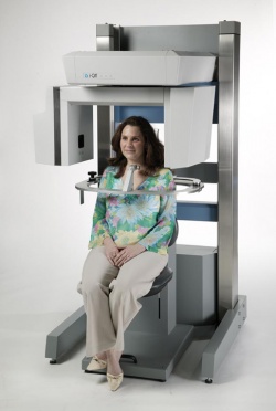
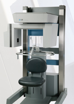
CBCT Cone Beam Computed Tomography (CBCT) scanners in dentistry have been in use in Europe since 1999. In the USA the FDA approved CBCT as a diagnostic imaging tool in dentistry. Since this, CBCT has emerged as one of the most useful tools in the diagnosis and treatment planning of complicated surgical procedures in dentistry. The compact size and relatively low radiation dose for the patient made CBCT ideally suited for imaging of craniofacial structures, including dental structures. CBCT has been widely used by oral and maxillofacial radiologists, oral surgeons, periodontists, prosthodontists, orthodontists and general dentists. Recently, UMKC SOD installed a CBCT unit, but this diagnostic tool had been used by dentists less than initially expected.
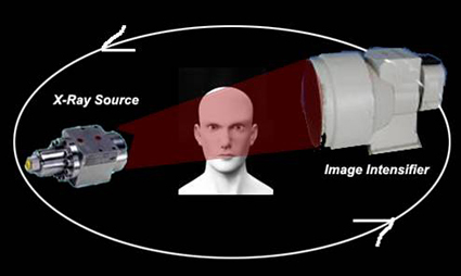
Cone Beam C.T: n Reduces patient’s exposure to 1/10th that of regular CT.n Have a cone beam X-ray Beam.n Images are displayed on monitor.n Images can be printed out on film, photographic paper or regular paper.
CBCT compare to MCTn 3-D Volume imaging similar to CT.n - Lower equipment cost.n - Simpler image acquisition.n Lower patient radiation dose.
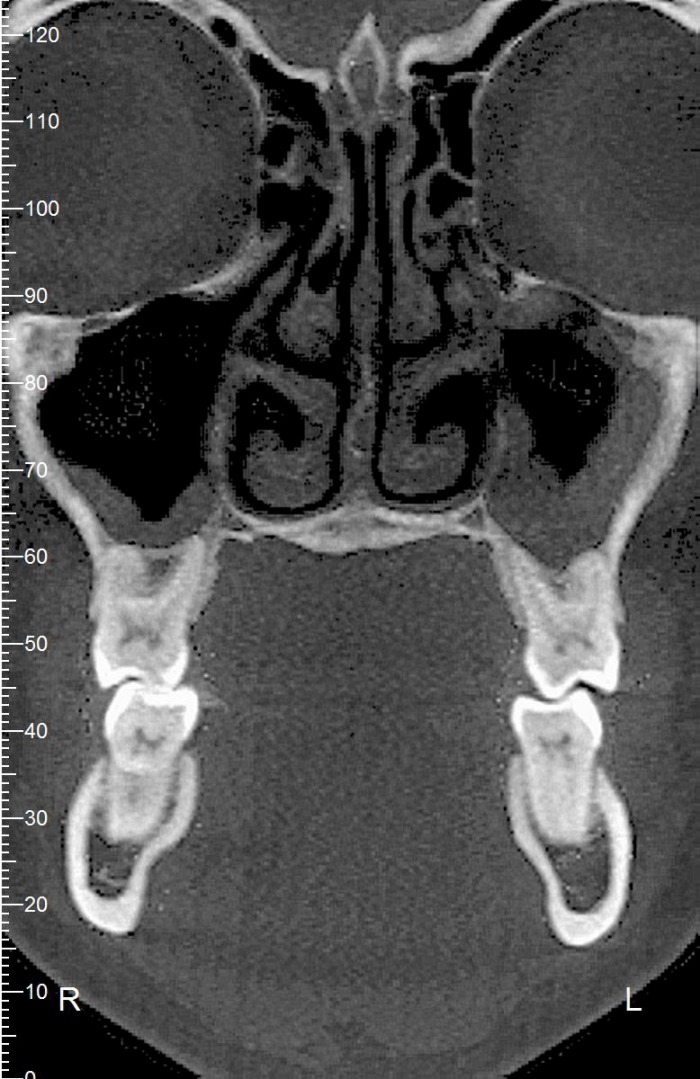
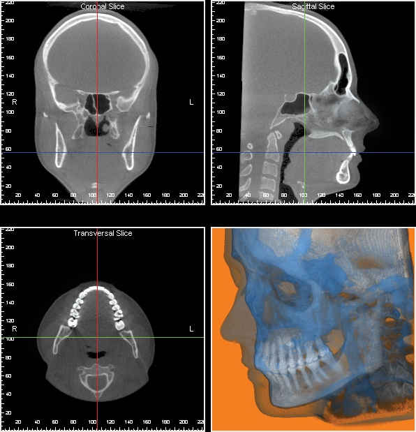
Assessment of pathology: The inherent high resolution of the CBCT ensures anatomic accuracy and assessment of all bony pathology in the maxillofacial region.
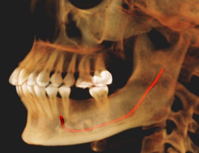
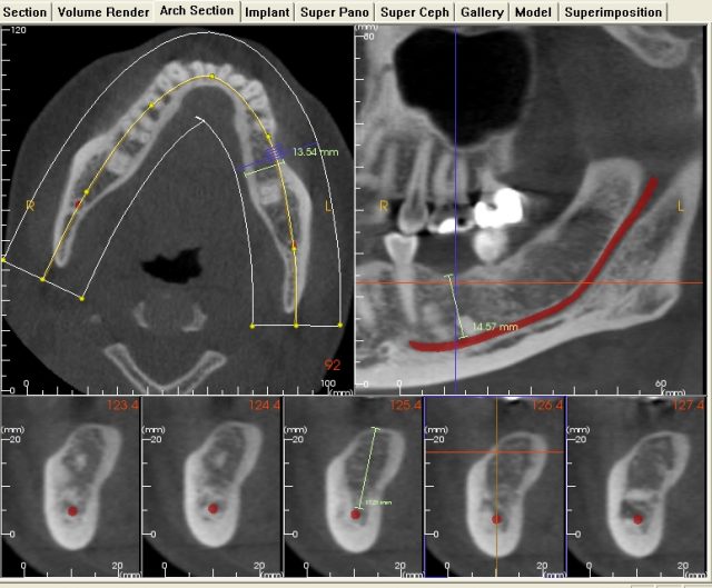
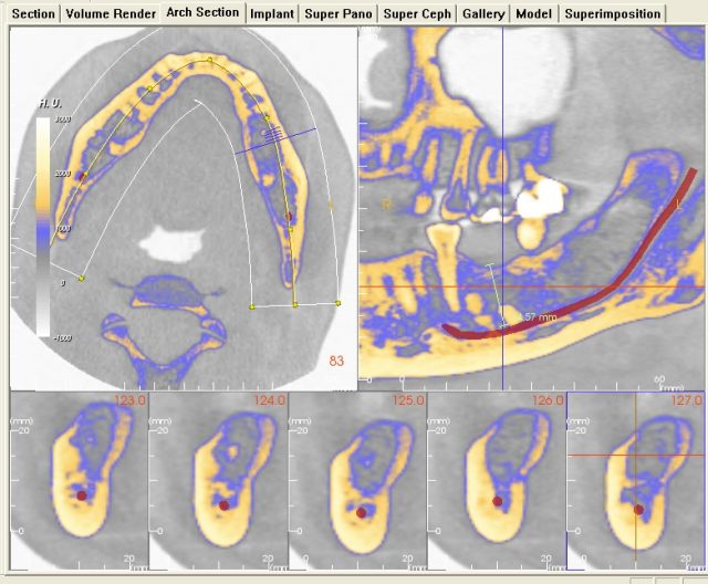
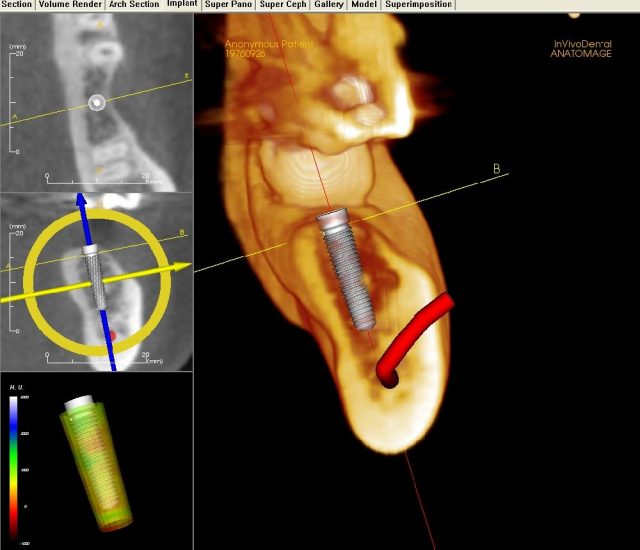
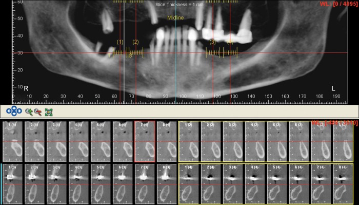
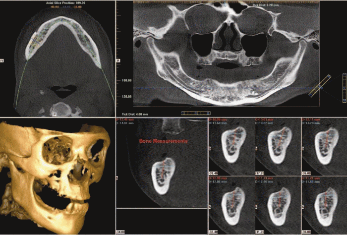
Dental Implants: One component of the successful placement of dental implants depends on the adequate assessment of the available height and width of alveolar bone at proposed implant sites. CBCT provides images at life-size which are familiar to surgeons (e.g. “panoramic”) and thin cuts at appropriate locations.
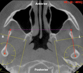
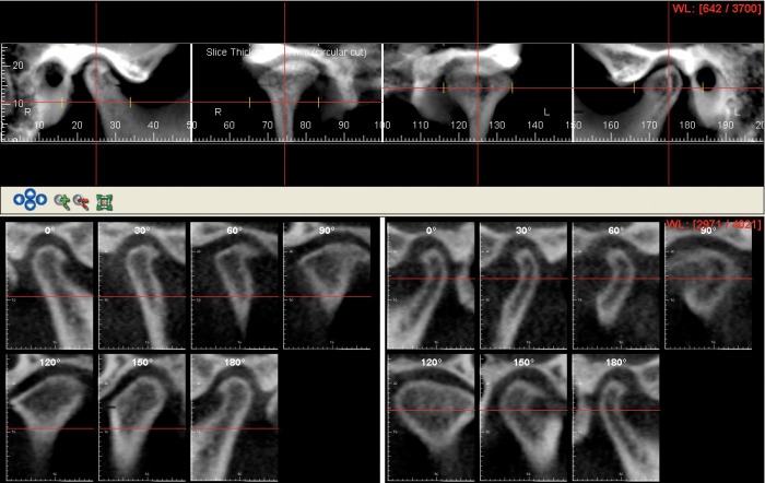
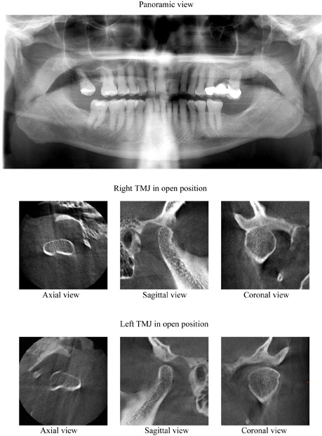
TMJ Disorders
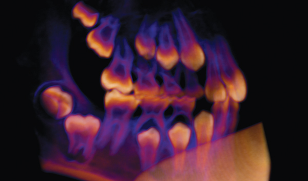
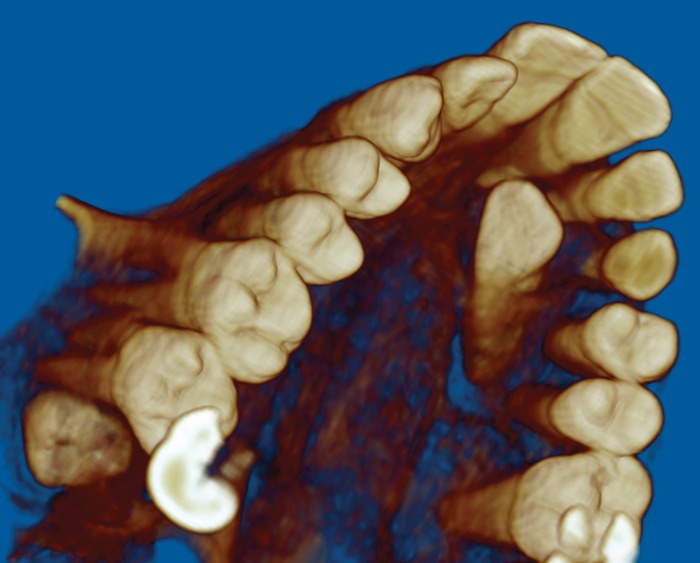
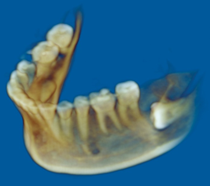
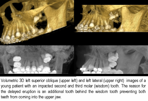
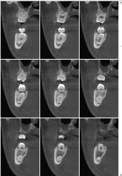
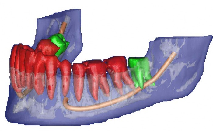
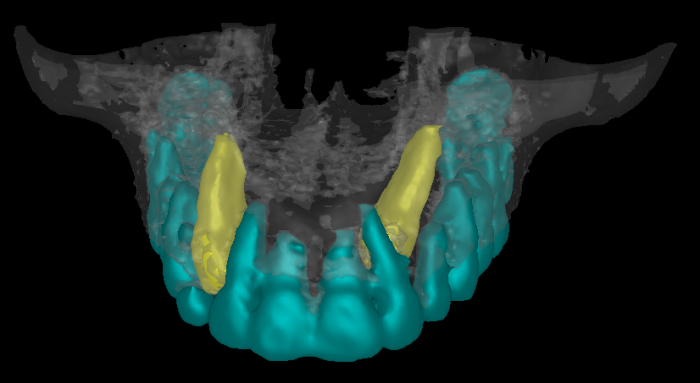
Localization of teeth: n The inherent high resolution of the CBCT ensures anatomic accuracy in the localization of impacted teeth as well as the assessment of root resorption or fusion (ankylosis).
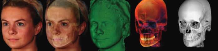
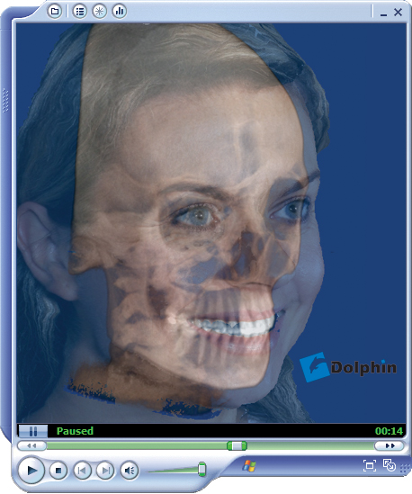
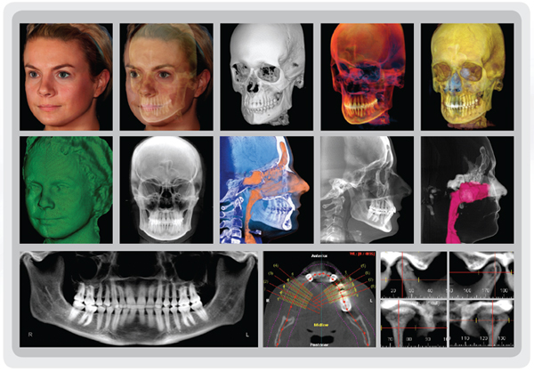
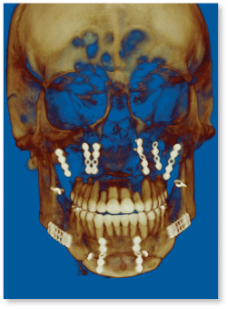
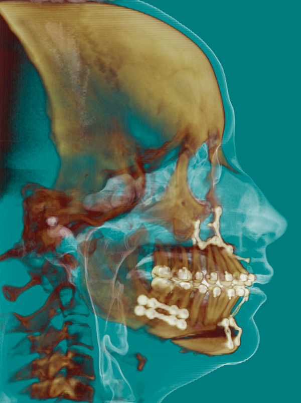
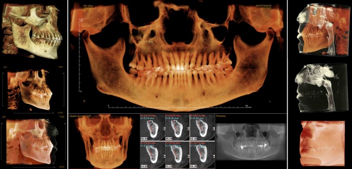
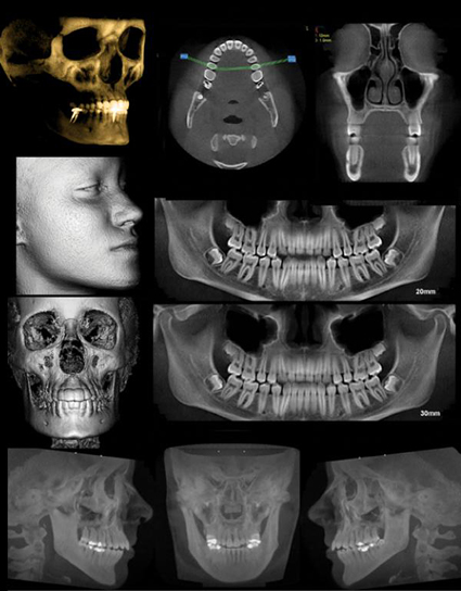
3D imaging / stereolithographic modeling: n Data from the CBCT can be reformatted to produce 3D images for the surgeon. This is important for patients requiring major reconstructive surgery. These images can be converted into models which assist the surgeon prior to the operation, reducing anesthetic cost and operating time. Technical advances in dentistry are occurring at an exponential rate. We have now arrived at the 3-D revolution with cone beam computerized tomography (CBCT). Since its arrival in the 21st century, 3-D diagnostic software and CBCT have set new standards in dental imaging.